Showing posts with label Tendon injury. Show all posts

Tuesday, 6 April 2010

Extensor Digitorum tenosynovitis


A guitarist came with complaints of left wrist pain and swelling a few days ago. She had been training vigourously and held the 'chords' with the fingers (left hand) and subsequently did her biceps and triceps curls using dumbbells. Soon after the episode she started having pain and noticed 'fullness' in the back (dorsum) of her wrist.

She was advised to do RICE treatment and apply the anti-inflammatory gel. She was also treated with Ultrasound therapy and isometric wrist exercises. After a few days, the symptoms subsided and she was given a step-by-step exercise program to prevent recurrence.

Read up more here.

Friday, 1 August 2008

Vastus lateralis tendinitis (tendinoses) and Iliotibial Band Syndrome


A forklift driver came this morning with complaints of left knee pain since the past 1 week with prolonged driving. His pain is worse at the end of the day and it was so bad that he could not sit or squat. He apparently felt better after massaging his thigh.

He had tenderness at the insertion of the vastus lateralis tendon, Iliotibial band (ITB)insertion and gluteus medius origin. He also had restriction of knee flexion (0-80 degrees) and had pain extending his knee against resistance.

As part of the treatment, he was given a day off, reassigned to other duties, told to do RICE treatment,given some stretching exercises for gluteal muscles, quadriceps and ITB and some NSAIDS for 5 days. He would also benefit from ultrasound, TENS, myofascial release and he had some taping done to restrict the pull on the vastus lateralis tendon.

Tuesday, 8 July 2008

Shock Wave Treatment for Achillis tendon Calcific Tendinitis

A young male hockey player came with chronic pain in his Achilles tendon several months ago. His earlier Ultrasound scan showed calcification of his Achilles tendon adjacent to the insertion to the calcaneal bone. He was given extracorporeal shock wave therapy(ESWT) (2000 pulses) weekly and felt much better after the second treatment. He went on to play the Kuala Lumpur Hockey League without any symptoms after 8 weeks rehabilitation and has been symptom-free since then.

ESWT has been approved for plantar fasciitis and has been found to be useful in treating calcific tendinitis.

I took a few photos of the treatment session.


Related websites:
International Society for Musculoskeletal Shockwave Therapy
FDA New Device Approval
EMS Website
Principles of Shockwave Treatment

Thursday, 22 May 2008

Achilles Tendon Paratenonitis

Mr Lim is a businessman who does regular brisk walking. He came 2 weeks ago with complaints of painful swelling in his back of his left ankle (Achillles Tendon). He started using a pair of leather shoes with a high heel tab which comes in contact with the Achilles tendon every time he walks.


He was examined and I found that had pain standing on his toes (ankle plantarflexion) but little pain stretching his calf (ankle dorsiflexion). He had severe tenderness, oedema and mild redness over the middle of his Achilles tendon.


I advised him to use a low-cut shoe, apply RICE treatment and Anti-Inflammatory gel (e.g. Reparil, Volteran Emulgel). His gait was otherwise normal and he did not have any calf muscle spasm. He would do well with a course Ultrasound treatment.

Friday, 25 April 2008

Extensor Retinaculum Injury and Extensor Digitorum Longus tenosynovitis


A male artistic gymnast was seen a few days ago with complaints of pain in the dorsum of the left foot after intensive training involving runs and jumps since 2 weeks earlier. He had difficulty standing on-toes, taking-off from jumps and landing.

After examining his ankle, I found that he had slightly reduced range of plantarflexion in the left ankle due to pain. There was tenderness and mild oedema of the extensor retinaculum and mild tenosynovitis of the Extensor Digitorum Longus tendons. However, resisted extension was pain free.


I did a simple taping to prevent extreme plantarflexion while enabling him to point his toes during his routine(Two figure of 6, and a double medial and lateral ankle locks and closed up the 'holes'). That seemed to do the trick and he should be back doing some training. However, he was told to refrain from sprinting or jumping to his maximal height as it may aggravate the injury. He was advised to undergo physiotherapy to reduce the symptoms and strengthen his lower limb.

Tuesday, 15 April 2008

Bilateral Peroneal Subluxation

The videos of a fencing athlete showing both feet plantarflexed and going into eversion to initiate subluxation of the peroneal tendons.

The left peroneal subluxation (suffered after a game of futsal) causes pain with sprinting and quick lunges hence limiting his performance in his sport. His rehabilitation may take up to 8 weeks before return to sport.


The right preoneal tendon partially subluxes but doesn't seem to give the athlete any problems as he is still able to plantarflex comfortably without pain.

Also see a previous post on peroneal subluxation here.

Friday, 28 September 2007

Right Biceps Tendon Rupture


Mr K was tugging onto the rope in a tug-of-war when he suddenly felt a snapping sensation with a sharp pain in his right arm a month ago. He thought he had torn his muscle but after a few weeks he seemed to have recovered fully except for the bulge in his right arm (see photo and compare with the normal left arm). He told me that he used to work out regularly at the gymnasium and hence had a much bigger arm. It looks as if it had shrunk significantly in size.

He only came to see me today and I examined his arm and found the 'popeye sign' and smaller biceps muscle with reduced power against resisted flexion. His forearm supination and pronation seemed as strong as the left. His long head of biceps was not palpable suggesting that it was ruptured completely. He only had mild pain with flexion of a fully extended elbow. He reminded me of an older case of a rugby player who had a similar biceps tendon rupture a few years earlier.

I referred him to an Orthopaedic Surgeon who suggested an MRI to detect the location of the edge of the long head. We agreed that he probably needed surgery if he wanted full function and active sports participation. If he was much an elderly patient, some physicians prefer a conservative non-surgical approach.

According to Klonz et al, ruptures of the long head of the biceps are commonly caused by degenerative changes within the tendon. Non-operative treatment gives good results, the loss of power regarding elbow flexion and supination amounts to only 8-21%. Refixation may be indicated for cosmetic reasons and offers a small but evident improvement of flexion and supination power. Deformity of the slipped muscle can be corrected effectively. Residual complaints after conservative treatment often result from associated subacromial problems.

Useful Links:-
Arthroscopic-assisted biceps tenodesis for ruptures of the long head of biceps brachii
Functional results after suture repair in ruptures of the long biceps tendon with special consideration of subacromial impingement
MRI or MR arthrography: a useful tool for evaluation of the biceps tendon rupture
Proximal and distal ruptures of the biceps brachii tendon

Wednesday, 1 August 2007

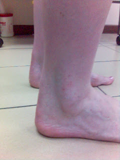
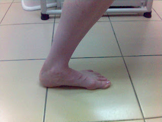
Tibialis Posterior Tendinopathy (Traction apophysitis)

Ms Chia is an 11 years old child who played basketball barefooted last week. She came with Right midfoot pain since a few days duration. She particularly mentioned that she had pain jumping and doing brisk walking. Her mother was worried as she thought it could be cancerous.

I examined her feet and found out that she was flat footed and had a prominent navicular tuberosity(insertion of Tibialis Posterior tendon) on both feet. She had tenderness at that spot but there were no obvious signs except that she had pain standing on her toes and resisting inversion (pushing her forefoot inward against my hand).

I explained to her mother that she would benefit by having good medial longitudinal arch support with orthoses. She could have taping of her foot to provide additional arch support. If symptoms persisted, an X-ray could be done to rule out avulsion fracture (tendon pulls off the tubercle from the bone) or other pathology.

She should recover swiftly with proper footwear, modified activity, isometric and theraband resistance exercises within 2-4 weeks.

Saturday, 28 July 2007

Peroneal Tendon Subluxation


Nadia (not her real name) was sub-elite gymnast when she came with sudden pain and snapping sensation in her right ankle after twisting her ankle during training 3 years ago. She was seen by another physician who diagnosed her condition and was treated conservatively with bracing for 1 month's duration followed by rehabilitation exercises for 8 weeks.

She had the snapping sensation when she stands on her toes ("On toes") and pointing her toes. I asked her to dorsiflex her ankle and evert the foot and her peroneal tendon would slip in front of the bony prominence (see photo). She only had mild pain everting her foot against resistance. As expected, there was a tendency for the opposite peroneal tendon to slip forward too due to increased laxity. Ultrasonography may be useful if we suspect a tendon tear.*

According to Ferran et al, the diagnosis and management plan are based on clinical evidence. Conservative management may be attempted in acute dislocations, and can be successful in up to 50% of patients, although there is a trend for operative management in athletes. Recurrent dislocations should be managed surgically.**

*J Ultrasound Med.2007; 26: 243-246
**Sports Med. 2006;36(10):839-46.

Tuesday, 17 July 2007

Flexor Hallucis Longus (FHL) Partial Rupture


Samson (not his real name) is a club soccer player who felt a sharp pain his right ankle injury during competition last Saturday. He felt pain in the inner aspect of the right ankle and could not 'push-off' to run or sprint after that. He used RICE treatment immediately after injury. The next morning he noticed a bluish discolouration of his hindfoot (see photo) due to dependent hematoma.

He saw me 2 days later and I had him dorsiflex his ankle and curl his toes downward against resistance (to strecth his FHL tendon). He grimaced in pain. He also had pain doing heel-lifts. I suspected a mild strain of his Tibialis Posterior tendon too.

He was seen by an experienced physiotherapist who told him he would be 'out-of-action' for at least a month. I told him that could be accurate but encouraged him to do some isometric exercises (without pain) and keep his fitness level by doing some aqua-jogging. He was prescribed some theraband resistance exercises and told to follow-up with his physiotherapist for electrotherapy.

For most practical purposes, he would need an ultrasound to evaluate the extent of the FHL partial rupture. Tendon partial ruptures may often require at least 12 weeks rehabilitation exercise followed by progressive return to play.

Tuesday, 8 May 2007

Achilles Tendinopathy


I remember treating 4 Korean women recreational athletes in the National Sports Institute a few years ago. They used to 'train' harder than some elite athletes often clocking up to 4 hours a day. A few of them suffered from Achilles tendon problems.

I had the opportunity to see another lady who was just as passionate about badminton. Ms Lee (not her real name) came with painful Achillles tendon (see photo) with localised swelling since 1-2 years duration. Her condition worsened over the past few months and she could not play badminton.


I found it rather interesting that she could not do a normal squat and had a thickened tendon an inch above the calcaneal bone attachment. It was certainly tender but more so along the inner side. As I palpated her calf muscles, she had spasm of her medial gastrocnemius causing her much pain stretching her calf muscle (see photo above)

She then told me that she had been playing on her toes thinking that her coach wanted her to literally 'play on her toes'! I was quite concerned that she was receiving various modalities of treatment but failed to undergo proper diagnostic or rehabilitative exercises. She was also an asthmatic on regular Inhaled Corticosteroid therapy provided by the Chest Physician (see Drug Saf, January 2005).

I quickly gave her some topical Arnica Comp gel to relieve the swelling, a mild Cox-2 selective NSAIDS (as she had gastric symptoms and could not tolerate non-specific NSAIDS) and some rehabilitation exercises. The rehabilitation exercises involved calf stretching and strengthening exercises, core stability exercises, hip and gluteal stretching, gluteal and hamstring strengthening and etc. I hope to see some progress in 2 weeks time but she will probably need at least 8-12 weeks before total recovery.

I would probably send her for an Ultrasound scan of her tendon or an MRI if she fails to recover adequately. If she was an elite athlete, she would have been scanned within the next few days to determine the prognosis. The ultrasound may reveal tears and degenerative changes e.g. calclfication.


If she continues to play without proper treatment she may have Achilles tendon rupture (see photo below). I would not recommend local corticosteroid injection for Achilles tendinopathy due to the risks of tendon rupture.

Athletes should also refrain from using Quinolone antibiotics due its toxicity on connective tissue increasing the risk of rupture especially in elderly. Arch Intern Med, August 2003.

Tuesday, 27 March 2007

Tibialis Posterior Tendinopathy


Ms Wang, a young lady executive shuffled slowly into my clinic. "I think I sprained my right ankle on the treadmill 2 days ago", she said. I was expecting a large swollen ankle but it was not that swollen. Then she pointed at the spot on the navicular tubercle (marked 'o' on the picture). She had a previous 'twist' of the same ankle several years earlier.

It sounded like a foot injury seen in my gymnasts, diving and dance sports athletes. She had mild pain when asked to flex and extend her ankle but she could not twist her foot outwards (eversion). She also had pain twisting her foot inwards (inversion) against the resistance of my palm. I palpated (felt with my fingers) the tibialis posterior tendon insertion on the navicular tubercle and she had severe tenderness.

Informed her that her tendon was strained but the deltoid ligament was spared. She needed to ice every 4 hourly (15 minutes/session), avoid high heeled shoes, possibly tape her ankle if she wanted to do more vigorous walking or running, have another look at her walking gait with her shoes, start some isometric exercises and progress to some theraband exercises. "I will see you after a week and expect the injury to heal within 3 to 4 weeks if you do your exercises"