Showing posts with label Rehabilitation. Show all posts

Wednesday, 11 June 2025

Foot bone contusion

 Getting a collision of the foot isn't a great thing to have. This chap limped in to see me with a slight swelling on his left foot. He thought it was fine as he was able to jog 6km in his sports shoes. However, he was concerned that he wasn't able to play football. His initial X-rays ruled out a fracture and the ultrasound scan ruled out tendon involvement. (One may resort to do an MRI if there is a high index of suspicion of a stress fracture especially if he has prodromal pain). We resorted to focal shockwave to sort out any bone oedema. He felt much better and was hopeful to play soon. Whatever it is, he would still need to have full pain-free function to execute all the football skills when he goes back to sports specific rehabilitation next week. 


Wednesday, 4 June 2025

Acromioclavicular Joint Arthrosis

 Had a young player come by with Right shoulder pain and restriction of overhead movement recently. His main complaint was he couldn't scratch his back unlike previously. Apparently he slept wrongly and woke up with the pain the next day. He had a Diclofenac sodium injection (Non-steroidal Anti-Inflammatory Drug) from a local doctor and felt better. However, there was some residual pain which lasted for 2 months. He was still able to play competitively but needed kinesiotaping. 

We did an ultrasound scan to find out that the had sclerotic changes and bursitis at his Right Acromioclavicular joint. Upon discussing the options of treatment, he decided that he would try focal shockwave treatment instead of intra-articular corticosteroid injection. 


 He left without the kinesiotape and with the possibility of full overhead movement and was able to scratch his back. Will see him again after 2 weeks to see how he is, but it will be highly unlikely that he will need another treatment so soon. 

Monday, 26 May 2025

Challenging injuries and chronic pain in professional footballers - is there a way out?

 As I was preparing for my talk for the AFC Medical Conference, I had the opportunity to get the help of a fellow researcher and statistician from Aspetar to evaluate the data of my shockwave work in footballers. We realised that some footballers do suffer from nagging injuries which lingered on after months. Some of them continue to play but some may seek regular recovery work or rehabilitation with their medical team. Being inside and outside of the dressing room where the players prepare, it is clear that at times they would prefer to have these problems sorted out. 


From 2015-2024, we noted that such footballers were referred with the following injuries for focal shockwave therapy. These injuries include Achilles tendinopathy, ankle impingement, knee focal cartilage defect, plantar fasciopathy, patella tendinopathy and foot fracture / stress injury. Details of the findings will be available in the lecture "Pursuit for return to play with focal shockwave in challenging football injuries" on 23rd July 2025.  


Wednesday, 21 May 2025

Shoulder myofascial pain syndrome

Ever though that computer bag is just too heavy? You might just be right. 

Had a chap walk in to relieve his misery which had been troubling him for a few days. We looked at his posture and he had a lower affected painful shoulder with an elevated other shoulder. The movements of both shoulders were not well coordinated
He felt some pain doing shoulder shrugs and retraction. He had pain on palpation of his trapezius and levator scapula. He was started on physiotherapy and underwent focal shockwave with joint mobilisation. 
He was given the advice to carry his bag on both shoulders, resume rhomboid and trapezius strengthening along with regular stretching exercises. Will see him again soon to review his progress. 

Sunday, 18 May 2025

Plantar fasciitis in 2025

Patients (even athletes) often come with heel pain (see photo), getting out of bed just as they start walking or after getting up from prolonged sitting. Walking barefoot makes things worse and going into a pair of comfortable shoes 👟 with a heel cushion or heel lift helps. I've had this on several occasions myself, but got better after doing frequent calf raises.


Sports medicine physicians have started to diagnose this both with physical examination and imaging (X-ray and Ultrasound scan) for better clarity. 

Clinically, the patient may have palpation pain at the bottom of the heel (inner part more common), and occasionally also at the back of the heel (Achilles tendon). The doctor would often examine the whole plantar fascia and calf for other signs of stiffness and pain. 


A quick prescription of silicone heel cup and taping by your physiotherapist would work wonders along with calf stretching and strengthening exercises. If pain persists, you could see your doctor who could prescribe further exercises, shockwave therapy or a local Platelet rich plasma or corticosteroid injection. You may seek further evaluation with your foot and ankle surgeon if 6-12 weeks of supervised physiotherapy does not resolve the pain. 

Saturday, 17 May 2025

Welcome home to Selangor FC - RGMC!

 A big welcome back to all readers of 'old-school' blogging! I've reclaimed this blog after more than a decade and decided to provide some lasting input on sports and musculoskeletal injuries.  


From my 13 year journey to the west (Doha, Qatar), I've decided to return home. You will see more about professional football, runners, swimmers, cyclists, musculoskeletal pain syndrome, and my favourite topic, shockwave therapy. Many thanks to Aspetar HospitalNational Sports InstituteNational Sports CouncilMASMISMSTAMSSM, and AFC for providing a platform of development for me. 

A big thank you for the management of Selangor Football Club for providing this avenue to continue to allow me to serve the footballers and members of the public. 

I am now available at RGMC in Section 5, Shah Alam, Selangor. Call us at 016-6251936 for appointments. The set up here has access for diagnostic, focal shockwave, injections, dietician, physiotherapy, strength and conditioning, etc. 


Friday, 16 March 2007

Ulna Neuritis in Cyclist

Mark is a 45 years old road cyclist with a mileage of 200 km per week. He came to see me 2 months ago with complaints of numbness and tingling sensation of his little finger and adjacent side of the hand. He mentioned that he was 'riding harder' than usual and had occasional wrist pain 2 weeks earlier as he 'felt good' and wanted to do more.

He was quite happy to lay off riding for 2 weeks and was given some isometric and resistance exercises with theraband. Subsequently, when I reviewed him again last month, he was slightly better. I gave him some neurotonics (vitamin B12) and allowed him to ride but advised against putting excessive pressure on the handlebars.

He came back last week without any symptoms and he said that he was riding better now that his wrists were stronger.

Tuesday, 13 March 2007

Plantar Fasciitis (Ouch!)


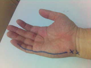
Brian is a 'recreational' badminton player who just returned to play after a 1 month celebration of Chinese New Year. For obvious reasons he wanted to get rid of some of the 'excess' weight that he gained. 2 days after play he started having severe hindfoot pain in the mornings. The pain subsides after walking for half and hour. He seemed to feel better walking in his leather shoes but playing badminton was a painful affair.

The pain was spot on the calcaneal attachment of the plantar fascia (see the mark 'X' on the photo). He also had tightness in the calf muscles and was flat footed. He had difficulty doing calf stretching. "I never did this before", he said.

We got him to wear a slipper with raised heel support (1/4"), a pair of preformed insoles for his flat foot, daily calf stretching when he got back from work, and ice-massage over the spot. He also saw a physiotherapist for myofascial release for his calf. Within 2 weeks he was back playing. Obviously, he was told to avoid playing to vigorously!

Monday, 12 March 2007

Low Backache


Ms Lim, a lady in her twenties came to see me this morning with complaints of low backache after waking up from sleep. She told me that she did not do any physical activity except the washing the corridor the previous day. However, she was able to sit with mild pain and did not have 'shooting' pain to her legs. She was previously seen by my colleague 1 month ago for the same problem but it resolved with some vitamin B12 supplementation and an analgesic.

She bent her back backward and forward and had moderate pain on the left side but her range of movement was full. She had some tenderness of the left quadratus lumborum muscle and the posterior superior iliac spine on both sides.

She was not keen on doing exercises or investigating further as she felt it was not a serious injury. I left her with a muscle relaxant, 5 days of moderate analgesics and advice to remain 'active'. She was told she could return to see me again if symptoms did not resolve as 50% of non-specific low back pain tends to resolve on their own within a week. I informed her that she probably should not use the corset (see picture attached) for longer than 2 weeks duration.

Wednesday, 28 February 2007

Medial Collateral Ruptured!

She was running down the hill slope when she landed her foot into a small hole and felt something snap and felt a sharp pain in the inner part of her left knee. She fell over and grimaced in pain. Her athlete quickly took her by the shoulders and helped her up to the clinic at the sports complex.

Elaine knew that her knee was in bad shape. She could hardly stand properly and the knee seemed to be twisted outwards. 10 years ago she had an injury to the same knee but she could still walk with little support.

"Looks like you've really torn your medial collateral ligament", I said to her. We quickly got her to ice the knee and prepared her file for physical examination. She had the full range of movement(ROM) which was quite 'pain-free' but she had severe tenderness and moderate swelling at the femoral attachment of the MCL. There was mobility of the leg when it was stressed outwards. Her tests for Anterior Cruciate and Posterior Cruciate ligaments were normal. She was lucky that there was no joint effusion (swelling in the joint due to accumulation of fluid e.g. joint fluid, blood, pus).

"Your MCL is complete torn clinically, but you should be back in 2-3 months time with rehabilitation if nothing else is torn", I said. She was given a functional knee brace and sent for an X-ray. Her X-rays came back normal except for the fluid shadow. We planned her for an MRI within the next 3 days to ensure that the other structures like meniscus and cruciate ligaments were not torn.

She was told to continue icing her knee every 4 hourly and focus on isometric exercises for her quadriceps and hamstrings for the next 1 week. Zul, the physiotherapist was quick to show her the rehabilitation chart. He told Elaine that she would need to progress in stages as she improves her strength, stability and function of her knees. he also planned hydrotherapy sessions for her to maintain her 'aerobic' fitness

Wednesday, 17 January 2007

Right Ankle Lateral Ligament Complex Sprain


An athlete's swollen right ankle is shown in the photo above. He had twisted his ankle while landing awkwardly from a jump in the morning. It did not swell up much as he had applied RICE treatment and had it wrapped with crepe bandage. (Note: Do not massage a new ankle injury as it would worsen the swelling and pain).

I examined his ankle and found that he had a partially torn ATFL ligament and lots of soft tissue swelling in the outer part of his ankle. He also had pain when he plantarflexed or inverted his foot. There was difficulty balancing on his right ankle due to the loss of sense of position and balance (proprioception).

He was able to walk with a slight limp due to the pain but he was advised to rest from footwork or excessive walking for a few days. He was given an option to use an Aircast ankle brace to provide additional support. After applying RICE therapy for 3 days, there was only minimal swelling and he could walk normally. Although, the foot looked almost normal, he still had to undergo rehabilitation exercises to strengthen and provide the normal proprioception in his ankle. It often takes up to 4-8 weeks for rehabilitation of an ankle lateral sprain.